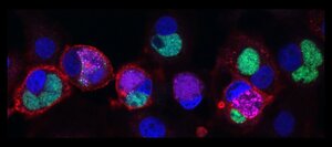
RO-37
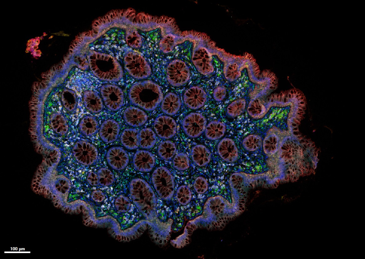
Colorful colon
This image shows a piece of human colon that has been fluorescently stained with a panel of markers for different types of immune cells. Additionally, dyes to visualize cell nuclei and an epithelial cell-cell adhesion protein were included. The tissue was then imaged on a multispectral slide scanner. Cross-sectioned colonic crypts appear as red circular structures while the underlying lamina propria contains a colorful mix of immune cells.by Mauro Esposito