RO-15
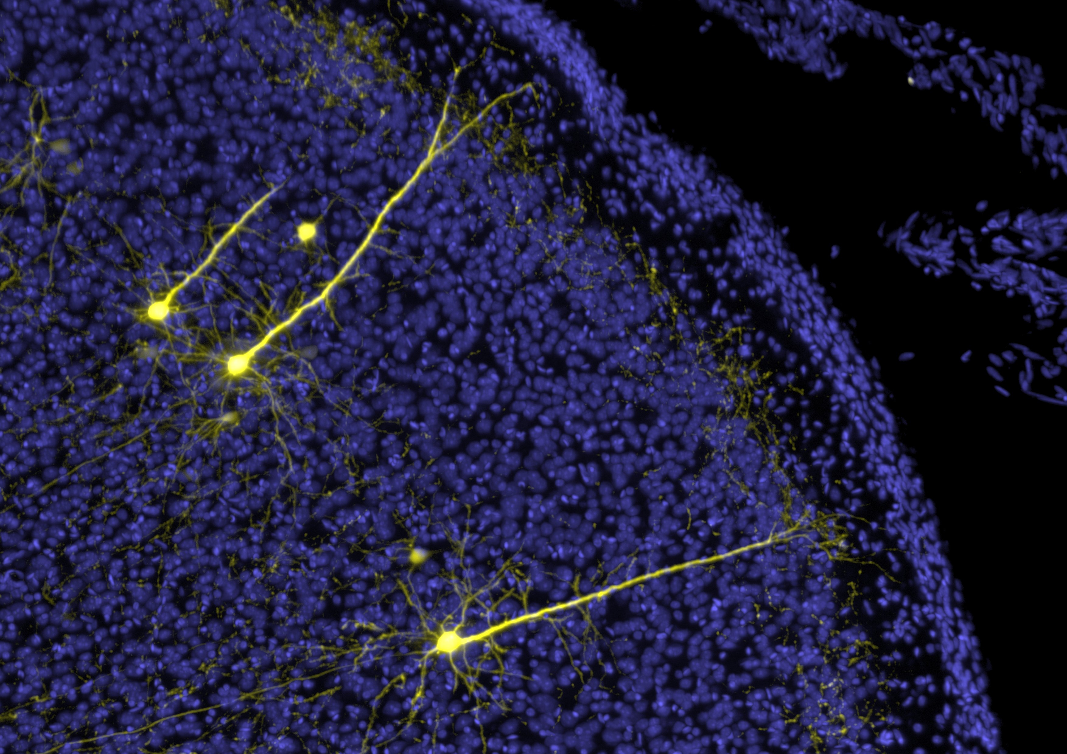
Pyramidal neurons (yellow) and cell nuclei (blue) in the mouse neocortex.
The image stack was acquired with a custom-built light-sheet microscope (Benchtop mesoSPIM) developed in Helmchen Lab, Brain Research Institute, UZH. The brain of a transgenic mouse was stained, cleared, and imaged in 3D at 20x magnification. Sparse staining and tissue transparency allows us to see the anatomy and distribution of neurons across the entire mouse brain.by Nikita Vladimirov